
THE JOURNAL OF ALTERNATIVE AND COMPLEMENTARY MEDICINE
Volume 11, Number 6, 2005, pp. 000–000
©
Mary Ann Liebert, Inc.
Evidence for Correlations Between Distant Intentionality
and Brain Function in Recipients:
A Functional Magnetic Resonance Imaging Analysis
JEANNE ACHTERBERG, Ph.D.,
1,2
KARIN COOKE, B.S., R.N.,
1
TODD RICHARDS, Ph.D.,
3
LEANNA STANDISH, N.D.,
4
LEILA KOZAK, M.S.,
4
and JAMES LAKE, M.D.
5
ABSTRACT
This study, using functional magnetic resonance imaging (fMRI) technology, demonstrated that distant in-
tentionality (DI), defined as sending thoughts at a distance, is correlated with an activation of certain brain func-
tions in the recipients. Eleven healers who espoused some form for connecting or healing at a distance were
recruited from the island of Hawaii. Each healer selected a person with whom they felt a special connection as
a recipient for DI. The recipient was placed in the MRI scanner and isolated from all forms of sensory contact
from the healer. The healers sent forms of DI that related to their own healing practices at random 2-minute
intervals that were unknown to the recipient. Significant differences between experimental (send) and control
(no send) procedures were found (
p
0.000127). Areas activated during the experimental procedures included
the anterior and middle cingulate area, precuneus, and frontal area. It was concluded that instructions to a healer
to make an intentional connection with a sensory isolated person can be correlated to changes in brain func-
tion of that individual.
1
INTRODUCTION
F
rom the beginnings of medical history, humans have
held a belief in a spiritual connection to others sepa-
rated from them at a distance. These beliefs have been held
as the basis for the efficacy of prayer, so-called energy heal-
ing, and the ability to heal others at a distance (“nonlocal
healing”). Despite the longevity of the concept, these
phenomena are largely dismissed by the advocates of the
biomedical model because they do not fit the current sci-
entific paradigm. The purpose of this study was to deter-
mine whether brain changes may be measured using fMRI
in the recipients of distant intentionality. In this paper, dis-
tant intentionality (DI) is used as a phrase that subsumes
prayer, energy healing, healing at a distance, spiritual heal-
ing, Therapeutic or Healing Touch, transpersonal imagery,
remote mental healing, and other practices based on puta-
tive connection in the absence of mechanisms of sensory
contact.
1
There is a growing interest in the scientific community
to study different forms of DI. In a recent publication sum-
marizing the current research on healing, at least 2200 pub-
lished reports on spiritual healing, prayer, energy medicine,
and mental intention effects were noted, as well as other ex-
amples of distant healing intentionality (DHI) or DI.
2
The
researchers noted the weak designs of many of the studies
1
Earl and Doris Bakken Foundation, North Hawaii Community Hospital, 67-1125 Mamalahoa Highway, Kamuela, HI.
2
Saybrook Graduate School, San Franciso, CA.
3
NeuroResearch Services, Kenmore, WA.
4
Bastyr University, School of Naturopathic Medicine, Kenmore, WA.
5
Department of Psychiatry, Stanford University Medical Center, Palo Alto, CA.
reviewed, concluding generally that the results merit further
study using sound methodology.
The neurophysiologic aspects of mystical, meditative,
or spiritual states have been studied with imaging tech-
nologies. Numerous studies of mystical or religious ex-
periences using single photon emission computed tomo-
graphic (SPECT) scans to capture brain function have been
reported.
3,4
Several of the studies showed reduced regional
brain metabolic activity in the posterior superior parietal
lobe during intense or peak religious moments. Among the
groups they studied were Tibetan meditators and Francis-
can nuns at prayer.
In a study of five individuals who had practiced Kun-
dalini yoga for at least 4 years, changes were found that oc-
curred in many areas associated with attention and control
of the autonomic nervous system (dorsolateral, prefrontal,
and parietal cortices, hippocampus, temporal lobe, and the
anterior cingulate cortex).
5
Still more relevant to the current study is the evidence of
correlative activity between the brain function of two indi-
viduals separated by distance, and in the absence of sensory
mechanisms of contact. What has been referred to as “ex-
trasensory induction” has been reported in 15 pairs of
monozygotic twins who were sensory-isolated from each
other and in separate rooms. In two of the 15 pairs, changes
in EEG alpha rhythms in one twin were observed simulta-
neously in the other.
6
A series of papers
7–9
reports several EEG studies show-
ing that a visually evoked potential in one member of a pair
of individuals who felt a close personal connection occurred
at above chance rates in the nonstimulated brain of the other
who was at a distance in an electromagnetically shielded
room. Although these studies were highly criticized because
of serious methodological issues, findings were later repli-
cated using appropriate statistical detection methods and im-
proved control conditions by two other independent labora-
tories using EEG technology.
10,11
An additional study that
employed a similar paradigm reported significantly corre-
lated fMRI signals between distant human brains.
12
Nineteen (19) studies replicating an apparent effect of in-
terconnectivity at a distance have been reported.
13
These
studies show above-chance correlations in electrodermal ac-
tivity (EDA), a measure of stress and arousal, between iso-
lated subjects. In their protocol, one subject was instructed
to randomly send anxiety provoking or relaxing images to
the other subject who was located in a distant room. Elec-
trodermal activity in the receiver subject was correlated
above chance, suggesting that the mental images of senders
influenced the state of arousal of receivers.
In summary, existing findings seem to suggest the posi-
tive effects of DI, the localization of brain areas activated
during prayerful or meditative states, and the correlation of
brain function between pairs of individuals. These data point
to the next logical step, which is to investigate the effect of
DI on the brain function of the recipient.
The research question investigated by this study is, “Is
there evidence for correlations between distant intentional-
ity and brain function in recipients of distant intentionality
who are tested using fMRI?”
METHODS
Subjects
Twenty-two (22) participants (11 pairs of healers and re-
cipients of DI) were recruited on the Big Island of Hawaii.
Healers were chosen who claimed to have the skills to com-
municate in some “nonlocal” form. In this first effort to doc-
ument the effect of DI, it was important to use participants
who already had training and experience in DI within their
traditions. There was no attempt to document their ability
to heal within the confines of this study. Often healers at-
tempt to heal illness of a psychological and spiritual nature,
and the typical medical records are of little use. To reiter-
ate, though, the study is not about healing per se, but whether
there is some correlation in the intention to connect at a dis-
tance with a person. The authors asked each healer to name
someone with whom they felt a bonded or close connection.
This decision was based on research cited earlier that indi-
cates close or bonded individuals may be more likely to
show correlated physiologic effects.
Inclusion criteria for the healers included:
1. Acknowledgment within their communities for their
healing abilities
2. Fulfilling cultural requirements for training, apprentice-
ship, and practice
3. Ability to name an individual with whom they claim a
special connection, who understands the goals of the ex-
periment and is willing to undergo an fMRI scan
4. A stated belief in their ability to turn on and off their
intentions within a time frame of approximately 2 to 4
minutes
The inclusion criteria for the receivers of DI included:
1. Being selected by the healer as someone with whom they
feel a close or empathic connection
2. Having the standard requirements for receiving an MRI
(no implanted devices or metal objects such as pace-
makers, joint pins) and no history of claustrophobia
3. Willingness to undergo an MRI scan of 34 minutes’ du-
ration and a postscan interview
Three (3) men and eight women with an age range of 46
to 71 participated as healers. The recipients of the healing
ranged in age from 44 to 61; and included three men and
eight women. On average, healers had been practicing their
healing traditions for 23 years.
ACHTERBERG ET AL.
2
The healers represented a variety of practices, including
Healing Touch (a practice of distant healing and laying on
of hands, conducted primarily by nurses trained in the
method); a traditional Hawaiian healing form termed
pule
that consists of prayer, chant, and song by a spiritual elder
or Kahuna; Peruvian shamanic healing; Reiki (a form of en-
ergy healing that may have ancient origins and was pur-
portedly rediscovered in the 19th century in Japan); vibra-
tion or sound healing, and three eclectic forms of DI that
did not fit into established traditions. Additionally, three of
the pairs represented a Chinese method of healing called
Qigong
, and all three trials were conducted by the same
qigong
master.
Procedures
The study protocol was approved by the Institutional Re-
view Board, University of Hawaii, John Burns School of
Medicine. The study was conducted in the Department of
Radiology, North Hawaii Community Hospital in Waimea,
Hawaii from August 2003 through July 2004.
Prescan
. Both members of each pair signed an informed
consent form and filled out a demographic questionnaire. A
semistructured interview was conducted with each healer
within 4 days before the scan to elicit information on his or
her DI practices. Then, the healers were given information
about their role, and the On (Send) and the Off (No Send)
procedures were described. Standardized instructions in-
cluded the information that they should try to connect with
the receiver during the On condition in ways that were pre-
scribed by their own DI practice. This was most frequently
described as sending energy, prayer, or good intentions, or
as thinking of the individual in the scanner and wishing for
them the highest good. All of the healers claimed that they
were not the cause of any healing effect, but rather were a
conduit for a spiritual or cosmic source. During the Off con-
ditions, they were instructed to take their attention away
from the person in the scanner.
The recipients of DI were instructed to relax as much as
possible in the scanner environment. They were provided
FMRI, DISTANT INTENTIONALITY AND BRAIN FUNCTION
3
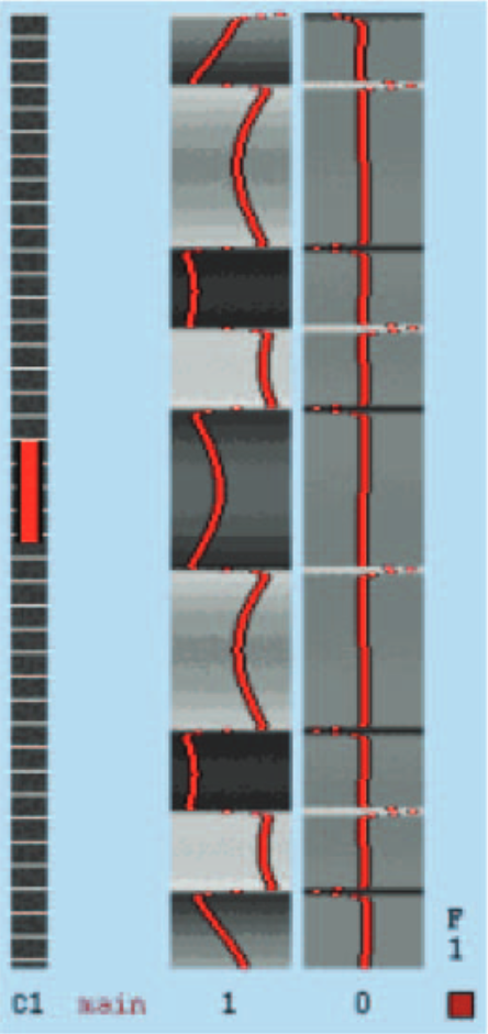
FIG. 1.
The design matrix used by the functional magnetic res-
onance imaging (fMRI) processing software to statistically analyze
the data. The vertical axis is in brain volumes (in this case 480 vol-
umes—one volume covers 3 seconds). The horizontal axis within
each column is related to the fMRI brain signal intensity. The red
line in the middle column indicates the model used in the General
Linear Model to fit the raw fMRI brain signals as a time series.
This timing is based on the healers’ timing, not the recipients of
DHI. This model was designed to account for possible habituation.
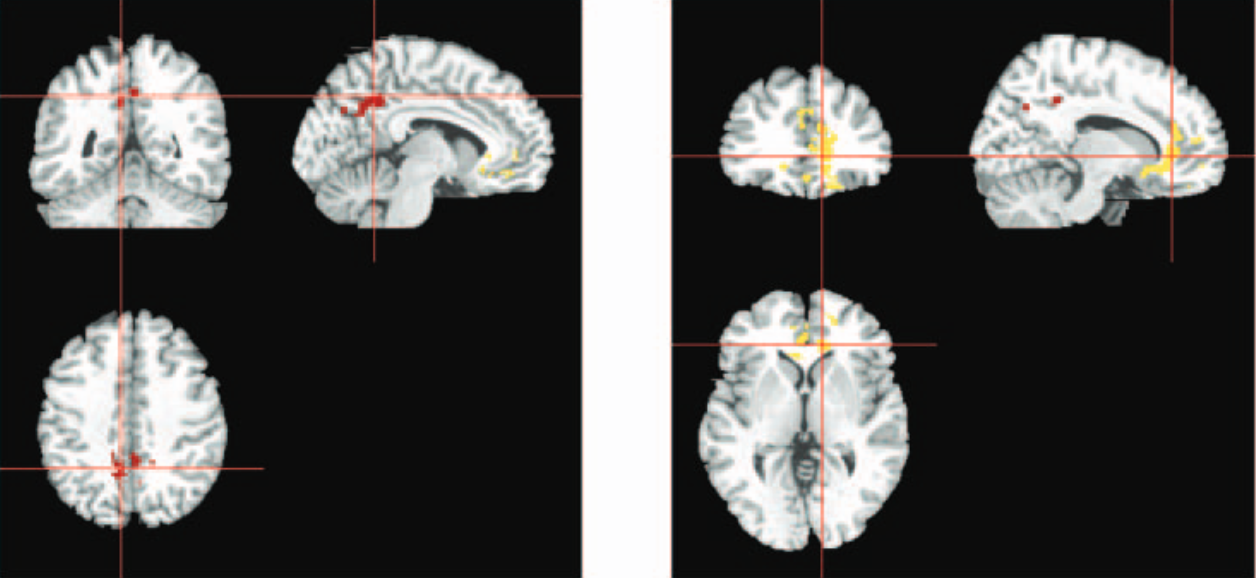
FIG. 2.
Group functional magnetic resonance imaging activation derived from 10 subjects where each individual subject was analyzed
with the On/Off paradigm of the healers. In the first scan on the left, the red areas indicate significant group activation in
the precuneus
and middle cingulate area. In the scan on the right, the yellow areas represent group activation in the anterior cingulate and
frontal regions.
QU1
4C
4C
with a call button and given instructions on using it if they
were distressed, had questions, or needed to stop the proce-
dure. However, no one used it for contact during the study
trials. They were made aware that the healers would be per-
forming DI. They were not provided with any information
about the timing of the On/Off conditions. Because the heal-
ers were not informed about the timing of the On/Off sig-
nals before the trials, they could not have coached their re-
ceivers before the scan.
Experimental conditions
. The healer was in the electro-
magnetically shielded control room and physically and op-
tically isolated from the receiver in the scanner. The radiol-
ogy technician, research nurse, and principal investigator
were also in the control room. During the course of the ex-
periment, the healer was verbally instructed by one of the
researchers with cues to start and stop the DI. The random
pattern of the twelve 2-minute intervals was determined
prior to the onset of the study using a coin toss.
A single randomized sequence that had an equal number
of on and off sessions was used for each session. The pat-
tern was
off, on, on, off, on, off, off, on, on, off, on, off
,
for a total of six 2-minute On periods and six 2-minute Off
periods. In three instances, it should be noted that the length
of the interval was 4 minutes because two of the On or Off
conditions occurred back to back. This pattern remained the
same for each healer. The total time the recipients of DI
were in the scanner was 34 minutes, which included a 10-
minute structural baseline of sagittal and transverse images.
During the time in the scanner, no physical or sensory con-
tact was made with the recipient by any member of the re-
search team.
Postscan
. The scan was followed by a semistructured in-
terview of both healer and receiver to elicit their subjective
experiences during the trial. Subjects in the study were paid
$100 for their participation.
FMRI Data Acquisition and Analysis
Structural and functional magnetic resonance imaging
was performed on a 1.5 Tesla MR imaging system (Siemens
Symphony Magnetom, Software Numaris/4, version Syngo
MR 2003BDHHS). The MR Symphony is up to specifi-
cations and is reliable as an MR unit. This has been veri-
fied through Siemens Medical Solutions Quality Assurance
procedures, which are monitored and verified through a
planned maintenance program and performed no less than
four times a year. Site personnel perform quality consis-
tency tests on a daily basis. In the facility where the scan-
ner is located the RF attenuation factors are Magnetic: 90dB
at 10.5 MHZ, Electric: 100dB at 10.5 MHZ. Blood oxygen
level dependent (BOLD) functional MRI scans were ac-
quired using a T2-weighted gradient echo version of the
echo-planar imaging (EPI) pulse sequence to identify re-
gional brain activation (transmits and receives radio waves,
pulses the magnetic field). Scanner protocols that are opti-
mized for measuring brain activation were used to maxi-
mize the BOLD response. Additional parameters of the
fMRI data acquisition include TR (time between pulse se-
quence cycles)
3, TE (time between the 90 degree pulse
plus the occurrence of the spin echo)
30 milliseconds;
slice thickness 6 mm, skip 1 mm; 64
64 acquisition ma-
trix; 21 slices positioned to cover the whole brain; and 408
brain volumes to cover the 24-minute acquisition period.
Four hundred and eight (408) brain volumes were collected
per subject.
Functional MRI scans were analyzed using the FSL soft-
ware program (Functional Magnetic Resonance Imaging of
the Brain, Software Library, Oxford Centre), which offers
robust corrections for false-positives, autocorrelation, mul-
tiple voxel testing comparison, and cluster size detection.
Analysis was carried out using FEAT FMRI Expert Analy-
sis Tool, Version 5.1, part of FSL. The following presta-
tistics processing was applied: motion correction using
MCFLIRT.
14
Time-series statistical analysis was carried
out using FILM (FMRIB’s improved Linear Model) with
local autocorrelation correction.
15
Z (Gaussianised T/F)
statistic images were thresholded using clusters determined
by Z greater than a cluster significance threshold of
p
0.01.
16–18
The corrected
p
value of 0.01 generally should
be found to be acceptable because it has been corrected
for multiple comparisons using the extent of cluster-size
algorithm.
Registration to anatomic images was carried out using
FLIRT. General Linear Model (GLM) regression was ap-
plied to generate statistical
p
value maps based on the con-
trast between the On versus the Off variables. The expected
response to changes in the healer/recipient protocol may be
equated to the expected response to stimulation paradigms
currently used in brain research. In these research para-
digms, the responses follow a hemodynamic delay curve.
The GLM regression can determine the extent to which the
observed receiver’s responses may be predicted by this
model. A goodness of fit statistic (
r
squared) indicates the
degree of fit between the hemodynamic model and the ac-
tual brain activity during the time course recorded. Both
positive and negative
coefficients can result from this
analysis.
The fMRI data were analyzed using the design matrix
shown in Figure 1.
The final step was to create the group maps from the in-
dividual fMRI analyses and coregister the group
z
-score map
to the MRIcro atlas (see ch2bet.hdr and aal.hdr from soft-
ware package www.psychology.nottingham.ac.uk/staff/cr1/
mricro.html) for the location and function of significant ar-
eas of activation. Then, software developed by one of the
authors (TR) was used to quantify the average
z
-score and
pixel activation counts within each of the 116 different brain
regions in the MRIcro atlas.
ACHTERBERG ET AL.
4
AU2
AU1
F1
Data were analyzed for both the intraindividual compar-
isons for the On/Off conditions (experimental versus con-
trol) and for the group effect as a whole during these pro-
cedures.
Ten subjects were included in the group analysis. One
subject was omitted because the fMRI analysis differed
slightly from the others in terms of number of volumes ob-
tained. Group and higher-level analysis were carried out us-
ing ordinary least squares (OLS) for simple, mixed effects.
Z (Gaussianized or normalized T/F) statistic images were
thresholded using clusters determined by
Z
2.3 and a (cor-
rected) cluster significance threshold of
p
0.01.
16,17,19
RESULTS
The FSL software produces a quantitative table of clus-
ter results that includes: cluster size, probability for each
cluster,
z
scores,
x y z
coordinates of the cluster in Talaraich
space and contrast of parameter estimates (see Table 1). If
a cluster is significant in a group analysis it means that there
were specific brain regions in which the combined subjects
had enough activation to raise the
z
score above the noise
level threshold.
In other words, if all of the subjects had random activa-
tion at different places in the brain, then there would be no
group activation. One of the two clusters was highly sta-
tistically significant (
p
0.000127). Significant areas of
apparent activation in the group analysis and total number
of pixels activated for the group are reported in Table 2. A
scan representing the group activation as a whole appears
in Figure 2.
DISCUSSION AND CONCLUSIONS
Group analysis revealed significant activation in several
areas of the brain, especially the anterior cingulate cortex,
frontal superior areas, and the precuneus. The authors’
anatomic definitions are correct if the Tzourio-Mazoyer at-
las is used.
20
It was produced as a segmentation of the MNI
atlas. This is available in MRIcro as ANALYZE files. The
conventionally ascribed functions of the cingulate cortex
are considered executive control, and decision making
at this level determines both verbal and motor responses.
The rostral anterior cingulate cortex area has been shown
to be activated during the height of opioid and placebo anal-
gesia response.
21
The frontal lobes are generally regarded
as modulating information processing, judgment, and deci-
sion making. Little is known about the function of the pre-
cuneus; however, it has been recently argued that it, along
with the anterior cingulate gyrus, may be a part of a neural
network that is involved in resting consciousness and self-
reflection.
22
Overall, the results show significant activation of brain
regions coincident with DI intervals. Even though the re-
sults of individual analyses and group analysis were sta-
tistically significant, the internal validity of these findings
is challenged on several fronts. First, from the data accu-
FMRI, DISTANT INTENTIONALITY AND BRAIN FUNCTION
5
T
ABLE
1. C
LUSTER
R
ESULTS FROM THE
G
ROUP
A
NALYSIS
Cluster -Log Max COG x COG y COG z
index Voxels P value 10(P) Z (mm) (mm) (mm) Mean COPE
2
1355 8.51E-09 8.07 3.82
9.07 40.5 1.42 17.5
1
475 0.00127 2.9 3.55
2.14
52.1 33.8 14.4
These are the Cluster results from the group analysis of combining the 10 subjects. There are two main clusters.
Column definitions: COGx
center of gravity of the cluster in Talaraich space x direction; COGy
center of gravity of the clus-
ter in Talaraich space y direction; COGz
center of gravity of the cluster in Talaraich space z direction; LOG10(P)
logarithmic
transformation of the
p
value; MaxZ
maximum zscore within the cluster; Mean COPE
contrast of parameter estimates (the
parameter estimated here is related to the change in brain signal intensity in comparing the on and off conditions in fMRI);
p
Value
probability of the null hypothesis that there is no significant activation which is based on cluster size and zscore values; Vo
xels
number of positive activation voxels within the cluster with zscore
2.4.
T
ABLE
2. S
UMMARY OF
A
REAS OF
S
TATISTICALLY
S
IGNIFICANT
A
CTIVATION
O
BSERVED IN THE
10 R
ECIPIENT
S
UBJECTS
Brain region Number of activation pixels
Frontal/superior/left 692
Frontal/superior/orbital/left 830
Frontal/mid/left 312
Frontal/mid/orbital/left 116
Frontal/inferior/orbital/left 335
Frontal/superior/medial/left 454
Frontal/superior/medial/right 122
Frontal/mid/orbital/right 368
Rectus/right 50
Rectus/left 438
Cingulum/anterior/right 866
Cingulum/anterior/left 1871
Cingulum/mid/right 163
Cingulum/mid/left 571
Olfactory/right 70
Olfactory/left 114
Precuneus/right 1466
Precuneus/left 1021
Caudate/left 139
T1
T2
F2
mulated it is not possible to establish causal factors for
the demonstrated effects. For example, three people (the
radiology technician, research nurse, and principal inves-
tigator) were in the control booth and aware of the timing
of the On and Off conditions. Given these facts, it is not
possible to know to what extent they influenced the find-
ings, even though they were not deliberately sending dis-
tant intentions. Second, because the study design used a
variety of healing traditions, one cannot know whether the
particular modality caused the effect or it was a function
of some unique and idiosyncratic interaction between
members of the pair. Finally, no independent measure of
the healer’s abilities is available. The healing traditions
represented are poorly researched, and the empiric evi-
dence for the prowess of any given practitioner is a mat-
ter of conjecture.
Given that there are no known biological processes that
can account for the significant effect of the DI protocol, the
results of this study may be interpreted as consistent with
the idea of entanglement in quantum mechanics theory.
23
Entanglement has been confirmed to occur between pho-
tons, and many have speculated that certain highly organized
macroscopic systems, including the brain, exhibit the prop-
erty of entanglement with other complex systems. In a re-
cent study evidence was found for nonlocal connections be-
tween separated preparations of human neurons.
24
These
findings, plus the current study correlating brain activity in
two sensory-isolated humans do not fit the classic model of
physics and can be interpreted as consistent with entangle-
ment at the macroscopic level.
Several future research directions are suggested, such as
replicating the present study using the same healers and re-
cipients; examining the importance of empathy or close re-
lationship by pairing healers with subjects who are unknown
to them; using a similar protocol to study possible relation-
ships between DI and healing in a sample of patients with
a particular medical diagnosis; studying possible group ef-
fects of several DI practitioners on a subject in the scanner
and scanning healers during the DI protocol with the goals
of identifying brain structures and functional brain changes
during the DI state.
In summary, these findings support previous research on
distant healing suggesting that human intentions may di-
rectly affect others in ways that are not entirely understood.
ACKNOWLEDGMENTS
The authors express deep appreciation to the Earl and
Doris Bakken Foundation, Kailua Kona, Hawaii, for the gen-
erous support of this research, and thank Steve Bauman
Ph.D. and Greg Hickok Ph.D. for their consultations. The
authors are grateful for the cooperation and facilities of
North Hawaii Community Hospital and for Roy Young’s
technical assistance.
REFERENCES
1. Schlitz M, Radin D, Malle B, et al. Distant healing intention:
Definitions and evolving guidelines for laboratory studies. Al-
tern Ther Health Med 2004;9(3suppl.):A31–43.
2. Jonas WB, Crawford CC, eds. Healing Intention and Energy
Medicine: Science, Research Methods and Clinical Implica-
tions. New York: Churchill Livingstone, 2003.
3. D’Aquili EG, Newberg AB. Mystical states and the experi-
ence of God: A model of the neuropsychological substrate. Zy-
gon: J Religion Sci 1993;28:177–200.
4. D’Aquili EG, Newberg AB. The neuropsychology of aesthetic,
spiritual, and mystical states. Zygon: J Religion Sci 2000;35(1):
39–51.
5. Lazar SW, Bush G, Gollub RL, et al. Functional brain map-
ping of the relaxation response and meditation. NeuroReport
2000;11(7):1–5.
6. Duane TD, Behrendt T. Extrasensory electroencephalographic
induction between identical twins. Science 1965;150(3694):
367.
7. Grinberg-Zylberbaum J, Ramos J. Patterns of interhemispheric
correlation during human communication. Int J Neurosci
1987;36:41–53.
8. Grinberg-Zylberbaum J, Delaflor M, Sanchez-Arellano ME,
et al. Human communication: the electrophysiological activ-
ity of the brain. Subtle Energies Energy Med 1993;3(3):
25–43.
9. Grinberg-Zylberbaum J, Delaflor M, Goswami A. The Ein-
stein/Podolsky/Rosen paradox in the brain: The transferred po-
tential. Physics Essays 1994;7(4):422–428.
10. Radin D. Event-related electroencephalographic correlations
between isolated human subjects. J Altern Complement Med
2004;10:315–323.
11. Wackermann J, Seiter C, Keibel H, et al. Correlations between
brain electrical activities of two spatially separated human sub-
jects. Neurosci Lett 2003;336:60–64.
12. Standish LJ, Johnson LC, Kozak L, et al. Evidence of corre-
lated functional magnetic resonance imaging signals between
distant human brains. Altern Ther Health Med 2003;9(1):128.
13. Schlitz M, Braud W. Distant intentionality and healing: As-
sessing the evidence. Altern Ther Health Med 1997;3(6):
62–73.
14. Jenkinson M, Smith SM. A global optimization method for ro-
bust affine registration of brain images. Med Image Anal
2001;5(2):143–156.
15. Woolrich MW, Ripley BD, Brady JM, et al. Temporal auto-
correlation in univariate linear modeling of fMRI data. Neu-
roImage 2001;14:6:1370–1386.
16. Worsley KJ, Evans AC, Marrett S, et al. A three-dimensional
statistical analysis for CBF activation studies in human brain.
J Cereb Blood Flow Metab 1992;12:900–918.
17. Friston KJ, Worsley KJ, Frakowiak RSJ, et al. Assessing the
significance of focal activations using their spatial extent. Hum
Brain Mapp 1994;1:214–220.
18. Jenkinson M, Bannister P, Brady M, et al. Improved opti-
mization for the robust and accurate linear registration and
motion correction of brain images. NeuroImage 2002;17L2:
825–841.
19. Forman SD, Cohen JD, Fitzgerald M, et al. Improved assess-
ment of significant activation in functional magnetic resonance
ACHTERBERG ET AL.
6
AU3
AU4
imaging (fMRI): Use of a cluster-size threshold. Magn Reson
Med 1995;33(5):636–47.
20. Tzourio-Mazoyer N, Landeau B, Papathanassiou D, et al. Au-
tomated anatomical labeling of activations in spm using a
macroscopic anatomical parcellation of the MNI MRI single
subject brain. Neuroimage 2002;15:273–289.
21. Petrovik P, Kalso E, Petterson KM, et al. Placebo and opioid
analgesia: Imaging a shared neuronal network. Science 2002;
295:728–737.
22. Kjaer TW, Nowak M, Lou HC. Reflective self-awareness and
conscious states: PET evidence for a common midline pari-
etofrontal core. Neuroimage 2002;17(2):1080–1086.
23. Einstein A, Podolsky B, Rosen N. Can quantum-mechanical
description of physical reality be considered complete? Phys
Rev 1935;47:777–780.
24. Pizzi R, Fantasia A, Gelain F, et al. Non-local correlation be-
tween human neural networks. In: Donkor E, Pirick AR,
Brandt HE, eds. Quantum Information and Computation II.
Proceedings of SPIE5436;2004:107–117.
Address reprint requests to:
Jeanne Achterberg, Ph.D.
North Hawaii Community Hospital
67-1125 Mamalahoa Highway
Kamuela, HI 95743
E-mail:
jeannieach@aol.com
FMRI, DISTANT INTENTIONALITY AND BRAIN FUNCTION
7
AU5